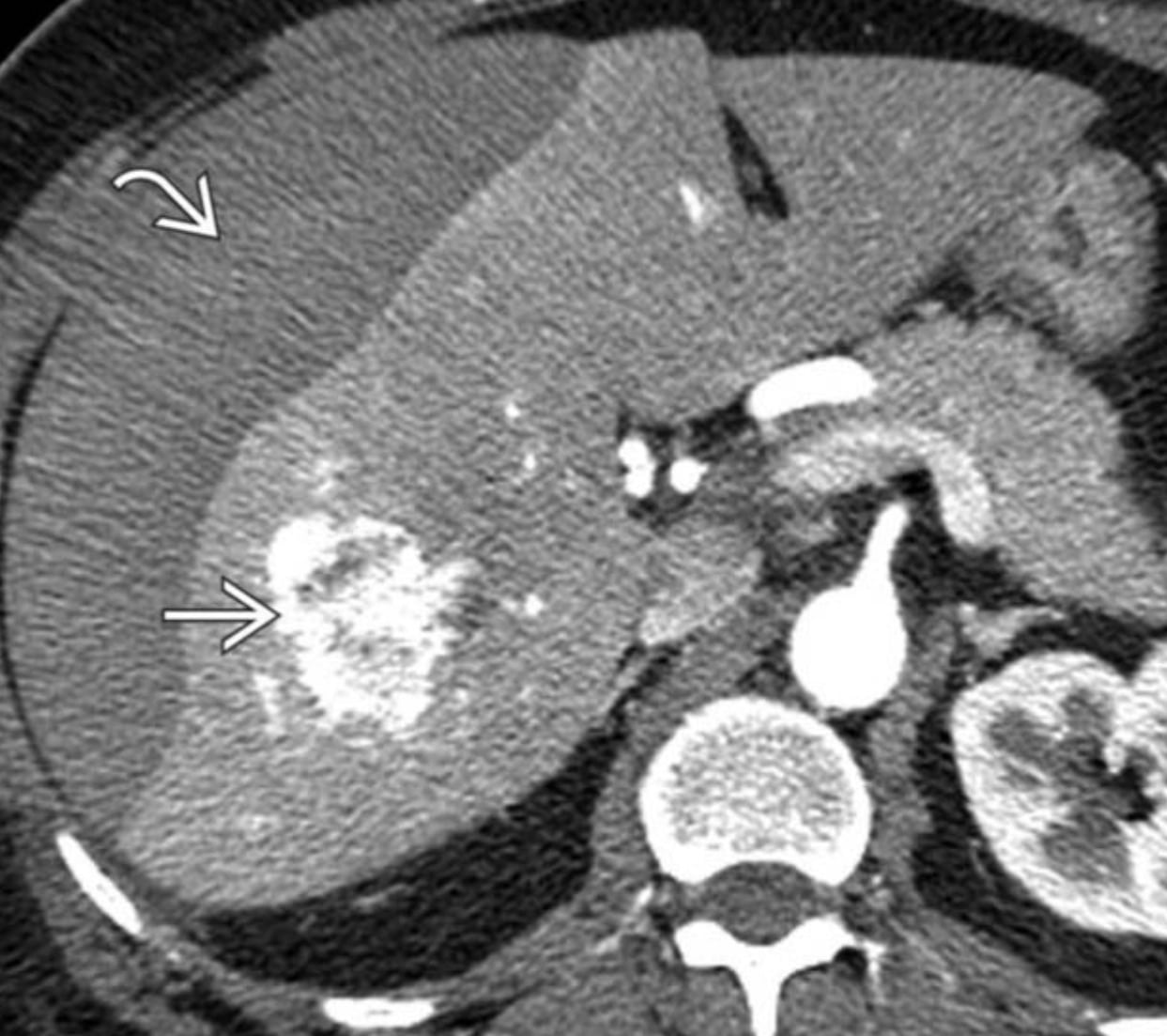
Hepatocellular adenomas (HCAs) are rare benign liver tumors that carry a risk of bleeding or cancer, particularly when larger than 5 cm.
Although classified into subtypes, management is mainly determined by tumor size and patient factors. Larger HCAs and those in men are typically removed, while smaller lesions can often be monitored. In practice, size is more important than subtype in guiding treatment decisions.
What is a Hepatic Adenoma?
Hepatocellular adenomas (HCA) are rare, benign (non-cancerous) liver tumors. While they usually don’t cause symptoms or require treatment, they can sometimes lead to complications such as:
- Bleeding (10%) – The tumor may rupture and cause internal bleeding.
- Malignant transformation (5%) – A small percentage of HCAs can turn into liver cancer (hepatocellular carcinoma, HCC).
Different Subtypes of Hepatic Adenomas
Not all HCAs are the same. They are classified into four subtypes based on imaging, pathology, and molecular markers:
- Inflammatory HCA (I-HCA) (40-50%) – More common in people with obesity, alcohol use, or metabolic syndrome. These tumors show signs of inflammation in the blood and liver tissue.
- HNF1A-mutated HCA (H-HCA) (30-40%) – These adenomas contain a lot of fat and almost never turn cancerous or bleed.
- Beta-catenin activated HCA (b-HCA) (10-15%) – This subtype has the highest risk of turning into cancer. Half of these tumors also have inflammatory features, making them difficult to identify with imaging alone.
- Unclassified HCA (U-HCA) (10-25%) – These adenomas don’t fit into any other category, and their cause is still unknown.
How Are HCAs Diagnosed?
Magnetic resonance imaging (MRI) is the best test to diagnose HCAs and distinguish them from similar liver lesions, such as focal nodular hyperplasia (FNH), which does not require treatment.
Each subtype has unique MRI features:
- I-HCA: Often appears bright on certain MRI images and may have a distinctive bright rim (the “atoll sign”).
- H-HCA: Contains fat, which is visible on specialized MRI sequences.
- b-HCA: No specific imaging pattern, making diagnosis more challenging.
When is an HCA Dangerous?
Risk of Bleeding
The main factor that determines the risk of bleeding is size:
- Tumors larger than 5 cm have a higher risk.
- Tumors near the liver’s edge (exophytic or close to the liver capsule) are more likely to rupture.
- I-HCA may have a slightly higher bleeding risk, but this does not change treatment decisions.
Risk of Cancer (HCC Transformation)
- Less than 5% of HCAs become cancerous.
- Larger tumors (>5 cm) have the highest risk.
- b-HCA and I-HCA subtypes are most likely to turn cancerous.
- H-HCA almost never becomes cancerous.
- Men have a higher risk of cancer from HCA than women, regardless of subtype.
Special Cases
Multiple HCAs (Adenomatosis)
- Defined as having more than 10 HCAs in the liver.
- Usually linked to metabolic conditions (e.g., glycogen storage disease, HNF1A-related diabetes).
- Not linked to hormone use (e.g., birth control pills, steroids).
- Treatment is based on size, just like solitary HCAs.
Pregnancy and HCA
- Estrogen (especially in the third trimester) can cause HCAs to grow or bleed.
- Women with HCAs larger than 5 cm should consider treatment before pregnancy to reduce this risk.
- If an HCA is found during pregnancy, surgery is safest in the second trimester if needed.
Do HCAs Need a Biopsy?
In most cases, biopsy is not necessary because MRI can provide enough information. A biopsy may only be considered if:
- The lesion is unclear (could be FNH or liver cancer).
- Knowing the subtype would change management (which is rare).
- The tumor is small and doesn’t meet the usual criteria for removal.
How Are HCAs Treated?
Lifestyle Changes
- Stopping oral contraceptives and anabolic steroids can help shrink HCAs.
- Weight loss and better metabolic control (in conditions like diabetes) may reduce tumor size.
Monitoring (For Tumors <5 cm)
- If the tumor is less than 5 cm, it can be monitored with annual ultrasound or MRI until menopause.
Surgery (For Tumors >5 cm or in Males)
Surgical removal is recommended for:
- Tumors larger than 5 cm (higher risk of bleeding or cancer).
- All HCAs in men (higher cancer risk).
Minimally invasive options like microwave ablation (MWA) may be considered for small or centrally located tumors.
Other Treatments
- Transarterial embolization (TAE) is used if there is sudden bleeding. It can also be considered as an alternative to surgery for non-cancerous HCAs.
Final Thoughts: Size Matters More Than Subtype
While identifying the subtype of HCA is useful for research, it rarely changes how the tumor is managed. Size is the most important factor in deciding treatment.
- HCAs smaller than 5 cm can often be monitored.
- HCAs larger than 5 cm or in men should be removed.
- MRI is the best tool for diagnosis and follow-up.
With careful monitoring and appropriate treatment, most people with HCA can be managed safely without complications.

